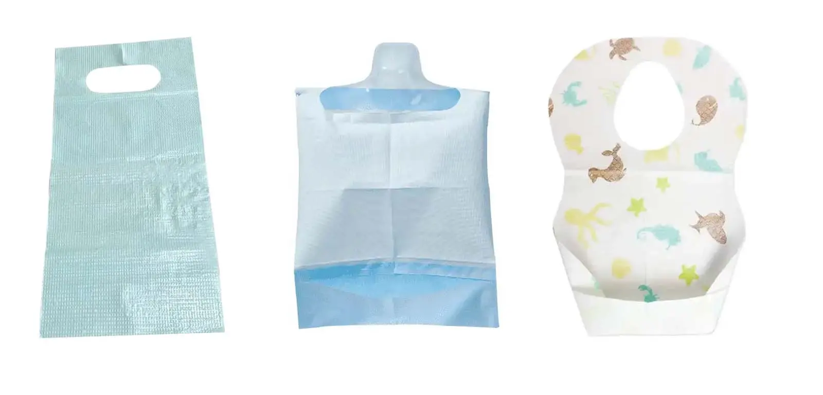
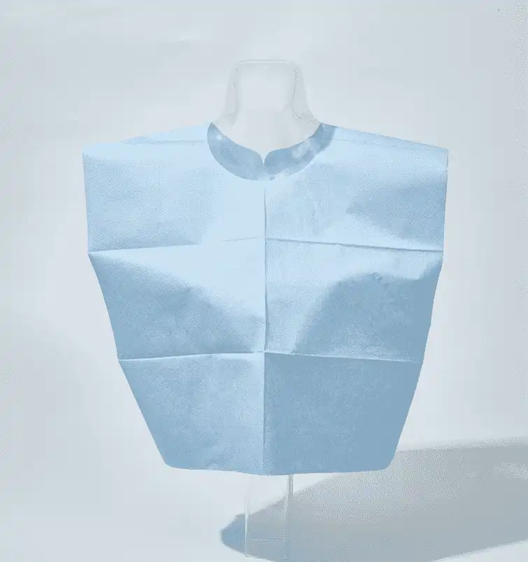
Are sterilization pouches the unsung heroes of dental clinics, silently ensuring the safety and well-being of patients and practitioners alike?
Exploring Cotton Sliver The Foundation Of Everyday Cotton Products
Cotton sliver is an intermediate stage in the process of turning raw cotton into the textiles we use daily. Understanding its role can provide valuable insights into both the textile industry and everyday products like medical cotton swabs and disposable cotton balls.
Beta-Carotene A Guide to Its Benefits and Sources
Beta-carotene is one of the carotenoids, an orange-yellow fat-soluble compound, which is the most ubiquitous and stable natural pigment in nature. Beta-carotene has high pharmacological and nutritional value, and has been widely used in medicine, beta carotene foods, food additives, cosmetics and animal feed industries.
Physical and chemical properties:
Beta-carotene is one of the most common isomers of carotene. The pure product is a reddish purple to dark red crystalline powder with a slight odor. The dilute solution is orange or yellow; Unstable to light, heat and oxygen, not acid resistant, but relatively stable to weak alkaline; Heavy metal ions, especially Fe3+, can make it fade. It has good coloring performance for oily food. It is a synthetic orange-yellow pigment that can be eaten, has vitamin activity, and is safer than other synthetic colorants.
Function:
1. β-carotene has independent physiological effects unrelated to vitamin A, which can enhance information transmission between cells;
2. Beta carotene is an antioxidant that cuts off the chain reaction and can eliminate toxic oxygen free radicals in the animal body;
3. Beta carotene can improve animal immunity, resist the invasion of bacteria and viruses, and improve the survival rate of animals;
4. Beta carotene can promote animal’s growth, improve reproductive performance, especially on female livestock, poultry;
5. Beta-carotene has a natural yellow or orange color and is also an effective colorant.
Practical application:
1. Improve product value, stimulate animal appetite: The purpose of adding colorants in feed is to increase the color of livestock, poultry and aquaculture animal products, such as the butter color of milk, the yolk and skin color of poultry eggs, and the feather color of birds. In addition, it can also change the color of the feed through the colorant to stimulate the appetite of livestock and poultry.
2. Improve animal immunity: Beta-carotene can exert antioxidant properties, thereby improving the animal’s immune capacity. Animals produce some highly active oxygen-containing molecules in the normal metabolic process, such as free radicals, hydrogen peroxide, etc. β-carotene can regulate the concentration of free radicals and hydrogen peroxide in cells, thereby improving the immunity of animals.
3. Enhance animal’s reproductive performance: Supplementation of a certain amount of beta-carotene to bulls can increase the number of sperm production and improve sperm motility. Beta-carotene is also very important for cows. A deficiency of beta vitamin in cows can lead to weak estrus, delayed ovulation, frequent follicular cysts, low conception rate, early pregnancy abortion, and in severe cases, can lead to reproductive disorders and placental arrest.
4. Promote the growth of Chinese soft-shelled turtle: β-carotene feed additive can enhance the disease resistance of turtle, promote the growth of turtle, improve meat quality, reduce feed cost, improve feed efficiency, and no residue, no drug resistance and other advantages, can make the produced turtle pollution-free, high nutritional value and good flavor, so it will be increasingly widely used.
Storage requirements:
Beta carotene supplementation should be kept in a cool and dry place, avoid direct sunlight and humidity, to avoid beta carotene weathering, deterioration and degradation, affecting the nutritional value and effect of feed.
The efficacy of Zinc L-Carnosine
Zinc carnosine (Zinc L-carnosine CAS No.: 107667-60-7) provides nutritional support for gastrointestinal mucosal healing. sinoway Zinc carnosine has been shown to help protect the gastric mucosa and promote gastrointestinal comfort. Studies have shown that zinc carnosine promotes healthy mucus production, exerts antioxidant properties, and inhibits the expression of cytokines that help protect the stomach and small intestine from injury. L-Glutamine promotes small intestinal cell function and mucosal integrity, further supporting gastrointestinal function and protecting tissue.
china-sinoway Zinc carnosine is a chelate of zinc with carnosine, not a simple mixture of carnosine and zinc. It selectively and persistently (it breaks down at a slower rate in the stomach) attaches to the gastrointestinal wall of the ulcer, like putting a "band-aid" on the skin, which helps to reduce inflammation of the ulcer ,heal. Carnosine zinc will eventually be slowly decomposed into L-carnosine and zinc, both of which are nutrients needed by the body, so the safety is good, and a reasonable dose will not burden the body.
What is UN3373 external or tertiary packing?
Badu(Xiamen) Technology Co., Ltd develops, manufactures and distributes a full line of solutions for the transport of biological substances of Category A (UN 2814 - UN 2900) and B (UN 3373), at room temperature, chilled or frozen.
The UN3373 specimen transport bag is a specific external or tertiary packing that is used for the safe transportation of biological substances, including clinical specimens, diagnostic samples, and other potentially infectious materials. These bags are designed to meet the requirements outlined by the United Nations (UN) for the transportation of biological substances, hence the code "UN3373."
When transporting UN3373 specimen transport bags, it is necessary to adhere to specific guidelines for packaging at different levels:
-
Primary packaging: This refers to the individual leak-proof specimen containers (e.g., tubes or vials) that hold the biological samples. These containers must meet the required standards for leak-proof and airtight sealing.
-
Secondary packaging: The UN3373 specimen transport bag itself serves as the secondary packaging. It is the bag specifically designed to enclose and contain the primary containers, providing an additional layer of protection. The bag should be leak-proof and resistant to damage during transportation.
-
Tertiary packaging: The tertiary packaging is the outer packaging that surrounds the UN3373 specimen transport bags. It is an extra layer of protection and containment to safeguard the specimens and ensure compliance with the transportation regulations. Tertiary packaging is usually required when shipping packages containing UN3373-specimen transport bags in bulk or for long-distance transportation.
The requirements for UN3373 tertiary packaging may vary depending on the specific regulations of the country or region. However, common considerations for tertiary packaging include:
-
Sturdy construction: The tertiary packaging should be sturdy enough to protect the UN3373-specimen transport bags from external impact or damage during transit.
-
Cushioning material: The use of appropriate cushioning materials, such as bubble wrap or foam, can provide additional protection against shocks and vibrations.
-
Sealing: The tertiary packaging should be secured with appropriate sealing methods, such as tapes or adhesive closures, to prevent accidental openings or spillage.
-
Clear labeling: The packaging should be clearly labeled with the appropriate UN3373 hazard symbol, indicating the presence of biological substances. Information about the shipper, receiver, and any necessary handling instructions should also be clearly displayed.
It is essential to consult and comply with the specific regulations and guidelines provided by the relevant authorities or organizations overseeing the transportation of biological substances, such as the International Air Transport Association (IATA) or national transport agencies. These regulations ensure the safe handling and transportation of UN3373 specimen transport bags and minimize the risks associated with the transportation of biological materials.
All our packaging complies with the recommendations and regulations in force by the ADR (road), RID (rail), ADN (inland waterways transport) IMGD (transportation by sea), ICAO (International Civil Aviation Organization) and IATA (international air transport).
The Functions of insulin cooler bag
The function of an insulin bag is to provide a portable and insulated storage solution for insulin and other diabetes supplies. Insulin needs to be kept at a specific temperature range to maintain its effectiveness, usually between 2°C and 8°C (36°F and 46°F) or as specified by the manufacturer.
An insulin bag is designed with insulation materials to help maintain the temperature stability of insulin during travel or daily routines. It helps protect insulin from extreme temperatures, whether it's hot or cold, which can negatively affect its potency. By keeping insulin within the recommended temperature range, an insulin bag helps ensure that it remains safe and effective for use in managing diabetes.
Insulin bags are often equipped with cooling elements such as gel packs or ice packs that can be frozen and inserted into the bag to provide additional temperature control. Some insulin bags also come with temperature monitoring features, like built-in thermometers or digital displays, to allow users to monitor and adjust the temperature inside the bag.
Overall, the primary function of an insulin bag is to provide a convenient and reliable way to store insulin while maintaining its temperature stability, allowing individuals with diabetes to carry their medication with them wherever they go.
What's The Principle of Immunofluorescence Assays?
Immunofluorescence (IF) is a powerful approach for getting insight into cellular structures and processes using microscopy. Specific proteins can be assessed for their expression and location, making immunofluorescence indispensable for scientists to solve many cell biological questions.
An immunofluorescence experiment is based on the following principal steps:
1. Specific antibodies bind to the protein of interest.
2. Fluorescent dyes are coupled to these immune complexes in order to visualize the protein of interest using microscopy.
It is distinguished between direct and indirect immunofluorescence. In direct immunofluorescence, the primary antibody is directly coupled to a fluorophore (also called fluorochrome), allowing for easy handling and quick visualization. In indirect immunofluorescence, a secondary fluorophore-coupled antibody, which specifically binds to the primary antibody, is used to visualize the structure of interest.
Although the second approach is more time-consuming than direct immunofluorescence, it has several big advantages, such as it is generally less expensive, because the secondary antibody can be used for different primary antibodies. In addition, several proteins can be specifically visualized in parallel in one single sample (multicolor immunofluorescence) by combining multiple primary antibodies with specific secondary antibodies—each of them labeled with a different fluorophore.
Nanjing Norman is a complete innovative diagnostic enterprise integrating research and sales. It has a comprehensive technical platform with complete instruments, reagents and reagent raw materials.
The company has successfully listed on the market with immunofluorescence analyzer FI-1000, FI-1200 and hundreds of testing items.
Nanjing Norman will continue to provide higher-quality products and services to global customers and partners! Welcome to send inquiry to info@nrmchina.com
Unveiling the Precision of Chemiluminescence Immunoassay in Modern Diagnostics
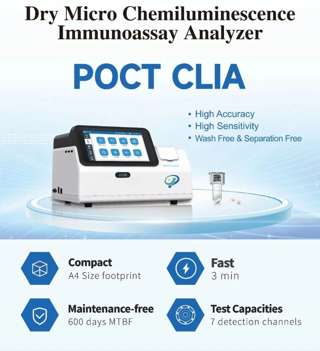
Welcome to our blog, where we delve into the fascinating realm of healthcare diagnostics and shine a light on one of the most innovative technologies in the field – Chemiluminescence Immunoassay (CLIA). In this article, we will unravel the mysteries behind CLIA, its applications, and the significant impact it has on modern medical diagnostics. Normanbio is professional Chemiluminescence Immunoassay analyzer and diagnostic equipment manufacturer.
What is Chemiluminescence Immunoassay (CLIA)?
Chemiluminescence Immunoassay, or CLIA, is a powerful and highly sensitive laboratory technique used for detecting the presence of specific molecules in biological samples. It combines the principles of immunoassay with the unique property of chemiluminescence – the emission of light resulting from a chemical reaction.
Key Components of CLIA:
Antibody-Antigen Interaction: CLIA relies on the specificity of antibodies to selectively bind to target antigens. This interaction forms the basis of the assay's accuracy.
Luminogenic Substrates: The magic happens when a luminogenic substrate is introduced. This substance reacts with an enzyme attached to the antibody-antigen complex, resulting in the emission of light.
Photodetectors: Specialized instruments capture and measure the emitted light, providing quantitative data about the concentration of the target molecule in the sample.
Applications of CLIA:
Clinical Diagnostics: CLIA is widely employed in clinical laboratories to diagnose various medical conditions, including infectious diseases, hormonal disorders, and autoimmune diseases.
Therapeutic Drug Monitoring: It plays a crucial role in monitoring drug levels in patients undergoing therapeutic treatments, ensuring optimal dosage and minimizing adverse effects.
Research and Development: CLIA is a valuable tool in pharmaceutical research, aiding in the development and testing of new drugs and therapies.
Advantages of CLIA:
High Sensitivity and Specificity: CLIA offers exceptional sensitivity, enabling the detection of low concentrations of target molecules with high precision.
Wide Dynamic Range: The technique can quantify a broad range of analyte concentrations, making it versatile for different diagnostic applications.
Automation and Throughput: Automated CLIA systems enhance efficiency, enabling high-throughput analysis, making it suitable for large-scale testing.
In conclusion, Chemiluminescence Immunoassay stands as a beacon of innovation in the field of diagnostics, illuminating new possibilities for accurate and efficient detection of various biomolecules. Its applications in clinical settings, research laboratories, and pharmaceutical development underscore its significance in advancing healthcare. As technology continues to evolve, CLIA is poised to play an even more prominent role in shaping the future of diagnostic medicine. Stay tuned for more updates on the latest advancements in healthcare technology on Norman's blog.
PFBN Proximal Femoral Bionic Intramedullary Nail Clinical Cases
Intertrochanteric fracture of femur is one of the common hip fractures in the elderly. The key of treatment are the strong fixation and early rehabilitation exercise, which can effectively reduce the occurrence of complications caused by bed rest and is of great importance to improve the recovery. However, the failure of traditional fracture internal fixation system is common in the treatment process. Recently, Liu Tao, director of the Department of Trauma and Orthopedics of Henan Provincial People's Hospital, took the lead in the first use of the international initiative and leading concept of the latest PFBN intramedullary nail fixation system in the treatment of intertrochanteric fracture successfully.
Case Information:
Wang, Female, 85 years old, 1 hour before the visit, she was injured in an accidental fall, presenting with pain in her right hip joint and limited movement. An axial position of the right hip joint was performed in the emergency department, and the emergency treatment was admitted to the hospital as "right intertrochanteric fracture".
Operation: closed reduction PFBN internal fixation
Postoperative: fracture reduction, good effect of internal fixator.
Checking Image:
Intraoperative Images:
Reduction:
Guide wire position:
Implantation:
Postoperative Images:
Minimally Invasive Treatment of Achilles Fracture with Arthroscopic Technique Using Type II Heel Nail-in-Nail System
Intra-articular calcaneal fractures are usually treated with traditional internal fixation involving a lateral widening incision followed by the use of calcaneal locking plates after resetting. While this method allows patients to return to work as soon as possible after the operation, it has several disadvantages. These include a large trauma caused by the L-shaped incision, the risk of skin necrosis, potential damage to the peroneal nerve during the operation, significant postoperative external adhesion, and limited mobility function. To avoid these complications, clinicians have started shifting towards minimally invasive internal fixation techniques.
Minimally invasive internal fixation has proven to be effective in treating patients with intra-articular fractures of the heel bone. It involves smaller incisions, utilizes X-ray fluoroscopy-assisted resetting, facilitates surgical operations, minimizes soft tissue stripping to protect the surrounding blood supply, and provides a favorable biological environment for soft tissue repair and fracture healing. This approach significantly reduces the incidence of adverse reactions and shortens the operation time. In recent years, ankle arthroscopy has also been increasingly used for diagnosing and treating intra- or extra-articular diseases of the foot and ankle. The application of arthroscopic technology in heel bone fractures, assisted by arthroscopy, brings new hope and direction to treatment. we are orthopedic implants manufacturer,website: https://www.doublemedicalgp.com